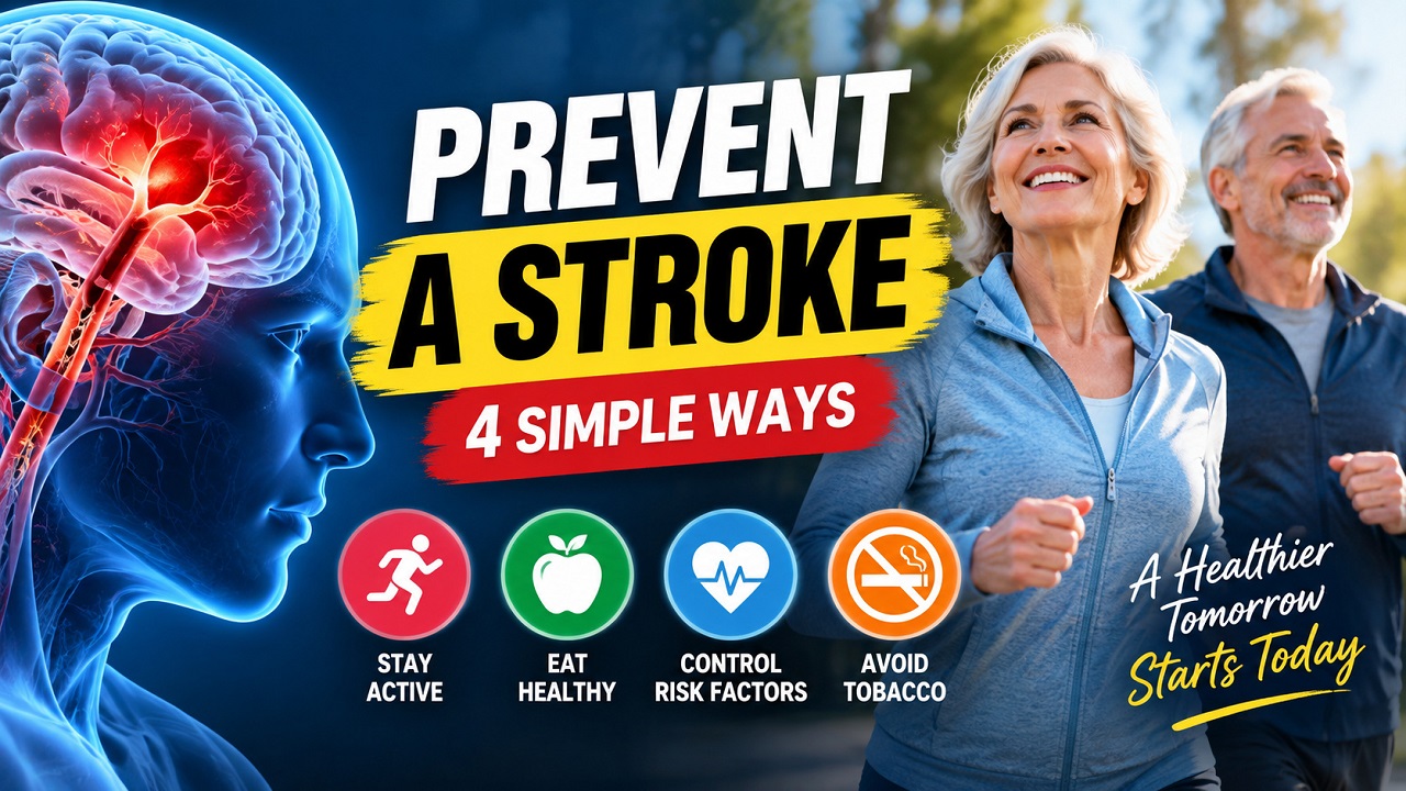
A cerebrovascular accident alters lives in seconds, yet clinical evidence demonstrates that up to 80% of premature vascular events remain entirely avoidable. When exploring how to prevent stroke, individuals confront dense medical literature that often obscures practical daily steps. Vascular integrity depends directly on systemic hemodynamic balance, cellular oxygenation, metabolic efficiency, and arterial elasticity. By addressing persistent hypertension, endothelial inflammation, and silent metabolic strain early, anyone can defend their neural pathways against progressive damage and secure vital long-term cognitive vitality.
TL;DR: Strokes occur when blood flow to brain tissue is obstructed by a thromboembolism or interrupted by vascular rupture. Long-term prevention focuses on aggressive control of high blood pressure, maintaining a dietary sodium limit below 2,300 mg daily (optimally 1,500 mg for hypertensive adults), eliminating tobacco use, and completing at least 150 minutes of moderate aerobic conditioning weekly. Chronic conditions including Type 2 diabetes, atrial fibrillation, and hyperlipidemia must be controlled through medical oversight and targeted lifestyle adjustments. Recognizing acute signs through the B.E. F.A.S.T. protocol ensures immediate medical rescue during critical vascular emergencies.
Quick Answer: Preventing a stroke requires keeping blood pressure below 120/80 mmHg, adopting a low-sodium Mediterranean or DASH eating pattern, exercising moderately for 30 minutes daily, quitting smoking, and eliminating excess alcohol consumption. Managing comorbid conditions such as dyslipidemia, diabetes mellitus, and cardiac arrhythmias through prescribed therapies drastically curtails clot formation and cerebral vessel damage.
A stroke is an acute neurological deficit caused by an abrupt focal vascular injury within the central nervous system, occurring when arterial blood supply to the brain is either obstructed by thrombosis or embolism (ischemic stroke) or interrupted by acute blood vessel rupture leaking directly into intracranial tissue (hemorrhagic stroke).
Primary Pathophysiology: Ischemic vs Hemorrhagic
Brain tissue relies on an uninterrupted flow of oxygenated blood. Interruption causes rapid ischemic cascades: ATP depletion occurs within minutes, followed by cellular depolarization, intracellular calcium accumulation, enzymatic breakdown, and irreversible neuronal apoptosis.
Systemic Vascular Damage
       │
       ├───────────────────────────────┐
       ▼                               ▼
Thrombosis / Embolism          Vessel Weakening / Rupture
       │                               │
       ▼                               ▼
Ischemic Stroke (87%)          Hemorrhagic Stroke (13%)
       │                               │
       └───────────────┬───────────────┘
                       ▼
             Neuronal Cell Death
Ischemic Stroke
Ischemic strokes account for roughly 87% of all cerebrovascular events. They develop through two primary routes:
- Thrombotic Events: Localized atherosclerotic plaque ruptures within major cerebral conduits (e.g., middle cerebral artery, internal carotid artery), triggering platelet aggregation that occludes the vessel lumen.
- Embolic Events:A traveling blood clot forms upstream—frequently inside the left cardiac atrium during atrial fibrillation—detaches, travels through arterial pathways, and lodges inside narrower distal cerebral capillaries.
Hemorrhagic Stroke
Hemorrhagic strokes result from structural vessel wall breakdown. Persistent mechanical shearing forces from uncontrolled hypertension weaken deep penetrating arteries, producing Charcot-Bouchard microaneurysms that eventually rupture. Alternatively, ruptured arteriovenous malformations (AVMs) or cerebral amyloid angiopathy (CAA) can trigger catastrophic intracerebral hemorrhage (ICH) or subarachnoid hemorrhage (SAH).
Clinical Summary:
Ischemic events stem from flow-blocking arterial clots; hemorrhagic events stem from vessel ruptures under elevated mechanical tension. Both deprive critical brain tissue of oxygen and glucose, requiring targeted medical interventions to preserve neural tissue.
Clinical Risk Factors: Modifiable vs Non-Modifiable
Comprehensive risk reduction requires separating fixed biological characteristics from modifiable systemic variables.
| Risk Factor Category | Clinical Indicator | Pathological Mechanism | Management Strategy |
| Modifiable | Hypertension | Endothelial shear stress and microaneurysm formation | Antihypertensive therapy, DASH diet, sodium limits |
| Modifiable | Atherogenic Dyslipidemia | ApoB/LDL-C oxidation driving atheroma maturation | HMG-CoA reductase inhibitors (statins), dietary fiber |
| Modifiable | Type 2 Diabetes | Glycation end-products inducing capillary stiffness | Metformin, GLP-1 receptor agonists, low glycemic intake |
| Modifiable | Atrial Fibrillation | Atrial blood stasis facilitating left atrial thrombi | Direct Oral Anticoagulants (DOACs), rate/rhythm control |
| Modifiable | Cigarette Smoking | Nicotine vasospasm, platelet hypercoagulability, carbon monoxide | Nicotine replacement therapy, behavioral cessation |
| Non-Modifiable | Age (>55 years) | Gradual arterial stiffening and microvascular rarefaction | Aggressive compensatory lifestyle control |
| Non-Modifiable | Genetic Predisposition | Monogenic disorders (CADASIL) or polygenic lipid traits | Serial biomarker screenings, early specialty surveillance |
| Non-Modifiable | Prior TIA or Stroke | Unstable vascular bed with high secondary recurrence rate | Antiplatelet regimens, dual therapy protocols |
Core Pillar 1: Blood Pressure and Vascular Health
Hypertension is the single most significant modifiable driver of stroke. Chronic arterial pressure damages smooth muscle layers within cranial vessels, accelerating atherosclerosis and provoking vessel collapse.
Elevated Arterial Pressure
       │
       ▼
Endothelial Shear Stress & Micro-Tears
       │
       ▼
Accelerated Plaque & Structural Wall Weakening
       │
       ▼
Lumen Occlusion OR Microaneurysm Rupture
Strategic Blood Pressure Management
- Target Optimal Ranges: Strive for systolic readings under 120 mmHg and diastolic measurements under 80 mmHg. Sustained readings above 130/80 mmHg require medical evaluation.
- Standardize Home Monitoring: Check arterial pressure twice daily (morning and evening) using a calibrated, automated upper-arm cuff. Avoid caffeine, exercise, and nicotine for 30 minutes prior to measurement.
- Ensure Pharmacotherapy Adherence: Prescribed angiotensin-converting enzyme (ACE) inhibitors, angiotensin receptor blockers (ARBs), calcium channel blockers, or thiazide diuretics should be taken consistently without sudden self-adjustments.
Clinical Summary:
Sustained blood pressure control reduces ischemic stroke danger by roughly 35% to 40%. Checking readings daily catches asymptomatic spikes before arterial damage progresses.
Core Pillar 2: Nutritional Interventions and Sodium Control
Diet directly modulates lipid subfractions, glycemic variability, and vascular endothelial function. High dietary sodium expands plasma volume and stiffens arterial walls, whereas adequate potassium intake helps counterbalance intravascular pressure.
Excess Sodium Intake (>6g/day)
       │
       ▼
Renal Water Retention & Fluid Volume Expansion
       │
       ▼
Increased Vascular Resistance & Shear Stress
       │
       ▼
Elevated Stroke Vulnerability
Evidence-Based Nutrition Guidelines
- Regulate Sodium Consumption:Adults should maintain salt intake below 6 grams (roughly 2,300 mg of sodium) per day, while individuals with verified hypertension benefit from keeping intake below 1,500 mg daily. Minimize packaged meals, preserved cold cuts, and restaurant sauces.
- Adopt Mediterranean and DASH Dietary Principles: Emphasize cold-pressed extra virgin olive oil, wild-caught fatty fish (sardines, salmon, mackerel), raw nuts, legumes, and cruciferous vegetables.
- Optimize Potassium-to-Sodium Balance: Incorporate potassium-rich foods such as avocados, spinach, sweet potatoes, and lentils to facilitate smooth muscle relaxation within the vasculature.
- Eliminate Industrial Trans Fats: Steer clear of partially hydrogenated oils and limit saturated animal fats to under 6% of total caloric intake to keep atherogenic LDL particles low.
Core Pillar 3: Physical Exercise and Metabolic Conditioning
Physical inactivity contributes substantially to metabolic dysfunction, insulin resistance, high resting heart rate, and chronic systemic inflammation. Sustained physical exertion triggers sheer-stress-mediated nitric oxide production, which dilates arterial walls and supports vascular elasticity.
Exercise Protocols for Stroke Prevention
- Moderate-Intensity Aerobic Regimen: Dedicate at least 150 minutes weekly to activities like brisk walking, cycling, or swimming at 50% to 70% of maximum heart rate.
- Vigorous Aerobic Conditioning: Alternatively, complete 75 minutes of high-intensity aerobic exercise, such as interval running or rowing, each week.
- Targeted Resistance Training: Integrate progressive resistance training twice weekly to enhance peripheral glucose disposal and support insulin sensitivity.
- Break Up Prolonged Sedentary Time: Take 3 to 5-minute walking breaks for every 45 minutes spent sitting to prevent transient vascular dysfunction and lower-extremity venous pooling.
Clinical Summary:
Consistent aerobic workouts support endothelial nitric oxide release, reduce resting vascular resistance, improve glycemic control, and stabilize body mass index.
Core Pillar 4: Smoking Cessation and Arterial Integrity
Inhaled tobacco smoke introduces thousands of toxic substances, including nicotine, acrolein, and carbon monoxide, that enter the bloodstream and damage arterial walls.
Tobacco Smoke Inhalation
       │
       ├─────────────────────────────────┐
       ▼                                 ▼
Endothelial Denudation & Spasm    Fibrinogen Elevation & Clotting
       │                                 │
       ▼                                 ▼
Accelerated Carotid Stenosis      Acute Thromboembolism Formation
The Pathological Cascade of Tobacco Smoke
- Accelerated Thrombogenesis: Inhaled toxins elevate fibrinogen levels and enhance platelet aggregation, significantly increasing clot formation within narrowed brain arteries.
- Lipid Peroxidation: Free radicals directly oxidize circulating LDL, quickening macrophage infiltration and plaque accumulation in the carotid bifurcation.
- Endothelial Dysregulation: Nicotine triggers sympathetic discharge, spiking acute blood pressure and causing microvascular constriction.
Cessation Milestones
- 24 Hours: Systemic carbon monoxide drops to baseline, raising arterial oxygen carrying capacity.
- 12 Months: Excess cardiovascular risk drops by roughly 50%.
- 5 to 15 Years: Overall stroke risk falls to match that of a lifelong non-smoker.
Core Pillar 5: Chronic Stress and Autonomic Regulation
Prolonged neuropsychological stress disrupts the hypothalamic-pituitary-adrenal (HPA) axis, flooding the cardiovascular system with cortisol and catecholamines. This sustained sympathetic state maintains elevated resting vascular resistance and increases inflammatory cytokines.
Evidence-Based Stress Mitigation Strategies
- Heart Rate Variability (HRV) Biofeedback: Engage in slow, paced diaphragmatic breathing (6 breaths per minute) for 10 minutes each day to enhance vagal tone and downregulate sympathetic activation.
- Sleep Hygiene: Maintain 7 to 9 hours of restorative sleep each night. Untreated obstructive sleep apnea causes nighttime hypoxemic surges and spikes early-morning stroke risk.
- Behavioral De-escalation:Research links chronic hostility, impatience, and intense anger with elevated stroke incidence. Cognitive behavioral strategies help reframe acute stressors and stabilize neuroendocrine patterns.
Clinical Summary:
Minimizing chronic stress reduces persistent sympathetic drive, prevents hypertensive surges, and curbs systemic arterial inflammation.
Community Insights: Real-World Experience and Misconceptions
Discussions across health forums and patient communities highlight practical concerns and common misunderstandings regarding stroke prevention.
Common Misconceptions
- Myth 1: “Strokes only happen to elderly people.”
- Clinical Reality: While incidence increases with age, roughly 10% to 15% of strokes occur in individuals aged 18 to 50. Carotid dissection, patent foramen ovale (PFO), thrombophilia, and premature atherosclerosis contribute to young-onset events.
- Myth 2: “Normal body weight guarantees vascular safety.”
- Clinical Reality: “Metabolically obese normal weight” (MONW) individuals often harbor visceral adiposity, severe dyslipidemia, and untreated hypertension despite an ordinary BMI reading.
- Myth 3: “Transient Ischemic Attack (TIA) symptoms that resolve require no follow-up.”
- Clinical Reality: A TIA is an urgent medical warning. Up to 20% of patients experiencing a TIA suffer a major stroke within 90 days, with the highest vulnerability occurring within the first 48 hours.
Common Pitfalls Reported by Patients
- Stopping prescribed antihypertensives or statins once readings normalize.
- Relying exclusively on over-the-counter supplements while ignoring high sodium intake.
- Assuming e-cigarettes and vaping present no vascular risks, despite evidence that nicotine continues to stiffen arterial walls.
Emergency Identification: The B.E. F.A.S.T. Protocol
Every minute of untreated large vessel occlusion results in the death of roughly 1.9 million neurons. Rapid recognition saves lives and preserves physical independence.
  B ── Balance: Sudden loss of balance or motor coordination
  E ── Eyes: Sudden loss of vision, partial field cut, or diplopia
  F ── Face: Asymmetrical facial droop or uneven smile
  A ── Arms: Unilateral arm or leg weakness, numbness, or drift
  S ── Speech: Slurred articulation, aphasia, or difficulty finding words
  T ── Time: Emergency! Note time of symptom onset and call emergency services immediately
Urgent Medical Directive:
If any single sign is present, immediately call regional emergency medical services. Never wait to see if symptoms improve, and avoid administering oral aspirin before a non-contrast brain CT scan has ruled out an intracranial bleed.
Prevention Protocols Across Age Groups
Tailoring interventions to specific stages of life ensures focused prevention.
| Stage of Life | Primary Vascular Threats | Core Screening Protocols | Primary Clinical Interventions |
| Young Adults (20–39) | Undiagnosed hypertension, recreational substance use, PFO, oral contraceptive hypercoagulability | Annual blood pressure checks, lipid profiles every 3 years | Tobacco cessation, regular cardiovascular workouts, balanced nutrition |
| Middle-Aged Adults (40–64) | Atherosclerosis progression, early metabolic syndrome, silent sleep apnea, early AFib | Annual vascular assessments, HbA1c tests, coronary artery calcium scoring, sleep evaluations | Strict sodium control, lipid-lowering therapies, weight management |
| Older Adults (65+) | Atrial fibrillation, severe carotid artery stenosis, arterial stiffness, cerebral amyloid angiopathy | Frequent pulse checks, carotid duplex ultrasound, continuous cardiac monitoring | Anticoagulation therapy when indicated, fall prevention, strict BP management |
Frequently Asked Questions
What are the earliest warning signs of an impending stroke?
The earliest signs often appear without warning and are best recognized using the B.E. F.A.S.T. framework. Key indicators include sudden loss of motor balance, unexplained acute visual loss or double vision, unilateral facial droop, arm weakness or numbness, and slurred speech or difficulty processing language. Some individuals experience a transient ischemic attack (TIA), where these signs emerge briefly for a few minutes before resolving completely. TIAs reflect temporary focal ischemia and demand urgent emergency assessment to forestall an impending full-scale stroke.
Can changing your diet reduce your stroke risk?
Evidence shows that dietary changes directly impact vascular health and significantly lower stroke risk.Adopting dietary models such as the Mediterranean or DASH patterns lowers systemic vascular resistance, reduces LDL oxidation, and regulates glycemic levels. Limiting sodium to under 2,300 mg daily (optimally 1,500 mg for hypertensive individuals) relieves fluid strain on the circulatory system. Emphasizing leafy green vegetables, soluble fibers, cold-pressed olive oils, and omega-3 fatty acids helps limit atheromatous plaque buildup within cerebral vessels.
How does high blood pressure cause a stroke?
Sustained hypertension forces blood through cerebral vessels at high pressure, causing mechanical micro-tears in the delicate endothelial lining. This chronic injury accelerates atherosclerosis by allowing oxidized lipids to build up in arterial walls, narrowing blood vessels and raising the likelihood of a localized clot. At the same time, prolonged pressure weakens the muscular walls of deep penetrating arteries, leading to microaneurysms that can rupture under strain and cause a hemorrhagic stroke.
Is stroke risk reversible after quitting smoking?
Vascular healing begins almost immediately after quitting smoking. Within 24 hours of cessation, carbon monoxide leaves the bloodstream, improving tissue oxygenation. Platelet aggregation normalizes rapidly, decreasing acute clot formation. Over twelve months of sustained cessation, excess cardiovascular risk drops by approximately 50%, and after 5 to 15 smoke-free years, overall stroke vulnerability approaches the baseline level of someone who has never smoked.
Preventing a stroke depends on consistent, deliberate vascular care rather than chance. By managing blood pressure, improving nutrition, incorporating regular aerobic activity, and eliminating tobacco use, you directly protect your cerebral arteries from progressive damage. Regular clinical screenings identify silent risk markers before symptoms emerge. Protecting long-term brain health and cognitive independence begins with manageable, deliberate steps taken every day.
Key Takeaways
- Control Blood Pressure: Maintain arterial readings below 120/80 mmHg to reduce ischemic risk by up to 40%.
- Cut Dietary Sodium:Keep total salt intake below 6 grams (2,300 mg sodium) daily to protect blood vessel walls.
- Stay Physically Active: Complete 150 minutes of moderate aerobic workouts each week to support vascular elasticity.
- Quit Tobacco Completely:Eliminating smoking prevents endothelial inflammation and avoids clot formation.
- Act Quickly on Symptoms: Use the B.E. F.A.S.T. protocol to spot symptoms early and contact emergency care without delay.
خدمات الرعاية الصحية والوقاية من السكتة الدماغية في أبوظبي
يقدم معهد الأعصاب التخصصي في كليفلاند كلينك أبوظبي رعاية طبية متطورة وفق أعلى المعايير العالمية في تشخيص وعلاج والوقاية من السكتات الدماغية وأمراض الأوعية الدموية العصبية. يضم المرفق نخبة من استشاريي الأعصاب وجراحي الأوعية، وتدعمه أحدث تقنيات التصوير بالرنين المغناطيسي والقسطرة الدماغية التداخلية لتقديم تدخل علاجي فوري وخطط وقائية متكاملة. يمكنكم التواصل مع كليفلاند كلينك أبوظبي لحجز موعد استشاري عبر زيارة الموقع الإلكتروني الرسمي clevelandclinicabudhabi.ae أو الاتصال عبر الهاتف المجاني 80082223 (من داخل الإمارات) أو 97180082223+ (دولياً)، ويقع المستشفى في شارع حمودة بن علي الظاهري، جزيرة المارية، أبوظبي، الإمارات العربية المتحدة.